

WHAT IS SPINA BIFIDA?
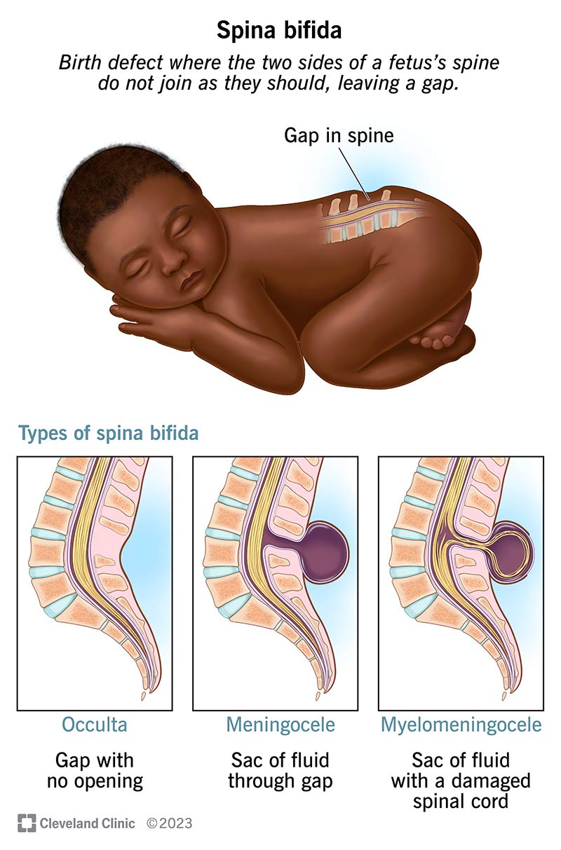
Spina Bifida is a type a neural tube disease that causes a developing fetus's spine to not properly close. When the neural tube does not close the backbone that protects the spinal cord and nerves are at risk for exposure causing damage. This damage can later lead to serious physical and intellectual disabilities to a child varying from a wide range of mild to severe. The severity of the disease depends on (1) the size and location of the opening and (2) how affected the spinal cord and nerves. Based on each of these characteristics Spina Bifida has been classified into 3 types; Spina Bifida Occulta (sounds like: o-cult-tuh), Meningocele (sounds like: ma-nin-jo-seal, and Myelomeningocele (sounds like: my-low-ma-nin-jo-seal). To read more about each of these please visit the Center for Disease Control'd (CDC) Spina Bifida Homepage.
Spina Bifida is a type a neural tube disease that causes a developing fetus's spine to not properly close. When the neural tube does not close the backbone that protects the spinal cord and nerves are at risk for exposure causing damage. This damage can later lead to serious physical and intellectual disabilities to a child varying from a wide range of mild to severe. The severity of the disease depends on (1) the size and location of the opening and (2) how affected the spinal cord and nerves. Based on each of these characteristics Spina Bifida has been classified into 3 types; Myelomeningocele (sounds like: my-low-ma-nin-jo-seal), Meningocele (sounds like: ma-nin-jo-seal, and Spina Bifida Occulta (sounds like: o-cult-tuh). To read more about each of these please visit the Center for Disease Control'd (CDC) Spina Bifida Homepage.
Lamin Keita at one month old.
(1) the size and location of the opening and (2) how affected the spinal cord and nerves. Based on each of these characteristics Spina Bifida has been classified into 3 types; Myelomeningocele (sounds like: my-low-ma-nin-jo-seal), Meningocele (sounds like: ma-nin-jo-seal, and Spina Bifida Occulta (sounds like: o-cult-tuh). To read more about each of these please visit the Center for Disease Control'd (CDC) Spina Bifida Homepage.
Diagnosis & Prevention
Spina Bifida can be diagnosed during pregnancy using various prenatal screening methods. These methods include blood test to screen for alpha-fetaprotein (AFP), ultrasound, and amniocentesis. However, in some cases Spina Bifida may not be detected until after pregnancy, in which, a number of imaging scans such as CT, X-ray, or MRI may be used. To learn more about the various test please visit the CDC's Spina Bifida Homepage.
Although, there is no clear cause to Spina Bifida, studies have shown that there are ways to reduce the risk. Our organization seeks to educate women who are pregnant on those methods in order to possibly prevent Spina Bifida such as 400 mcg of daily folic acid consumption post and during pregnancy.
Source: Center for Disease & Prevention: What is Spina Bifida? Accessed 7/17/2020
WHAT IS HYDROCEPHALUS?
Hydrocephalus, a complex neurological condition, poses significant challenges for healthcare professionals, particularly nurses, in providing specialized care to individuals affected by this condition. Characterized by the abnormal accumulation of cerebrospinal fluid within the brain, hydrocephalus can result in elevated intracranial pressure, leading to various neurological impairments.
Nurses play a crucial role in the multidisciplinary team, actively contributing to the assessment, management, and support of patients with hydrocephalus throughout their healthcare journey.
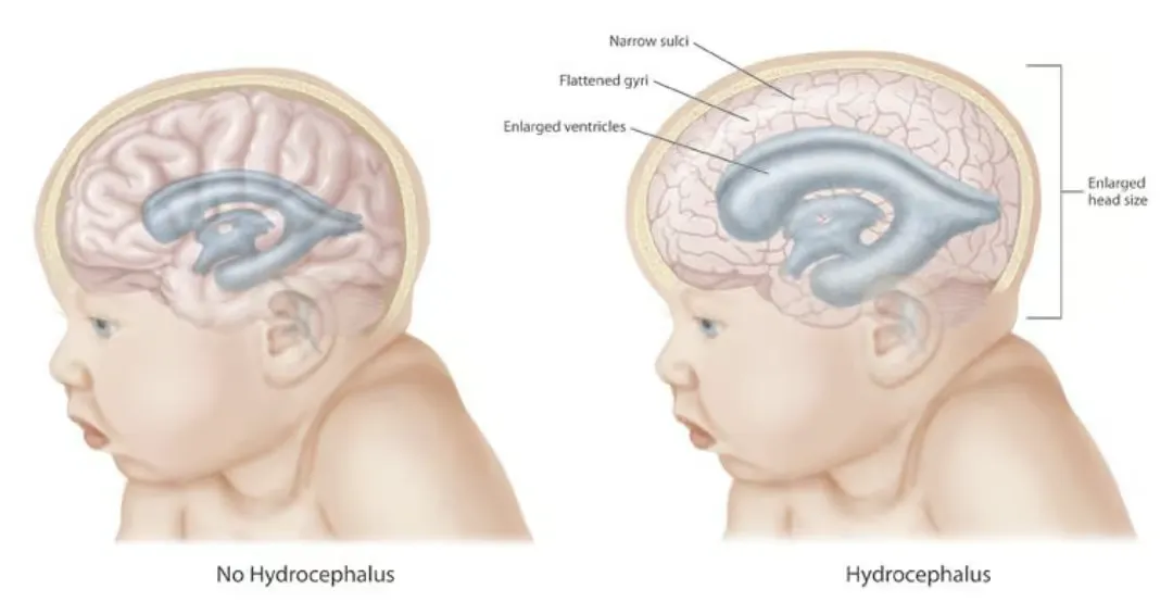
The term hydrocephalus is derived from two words: “hydro,” meaning water, and “cephalus,” referring to the head. Hydrocephalus is a neurological condition characterized by the abnormal accumulation of cerebrospinal fluid (CSF) within the brain’s ventricles, leading to an increase in intracranial pressure. This condition can occur at any age and may result from various underlying causes, such as congenital malformations, infections, tumors, or traumatic brain injuries. This condition could also be termed a hydrodynamic CSF disorder.
There are two types of hydrocephalus: noncommunicating and communicating. In the noncommunicating type of congenital hydrocephalus, an obstruction occurs in the free circulation of CSF. On the other hand, in the communicating type of hydrocephalus, no obstruction of the free flow of the CSF exists between the ventricles and the spinal theca; rather, the condition is caused by defective absorption of CSF, thus causing increased pressure on the brain or spinal cord.
Diagnosis and Medical Management

To diagnose hydrocephalus, medical professionals use a variety of imaging techniques, starting with CT scans to assess ventricle size and MRI to identify specific structural causes like tumors or malformations. In infants, ultrasonography is performed through the anterior fontanelle to monitor for hemorrhages or progressive fluid buildup. Skull radiography serves a dual purpose by detecting long-term pressure effects on the bone, such as a "beaten copper" appearance, and verifying the physical placement of shunt hardware after surgery.
More advanced diagnostic tools include Diffusion Tensor Imaging (DTI), which reveals microscopic white matter changes invisible to standard scans, and MRI cine, which measures the stroke volume of cerebrospinal fluid flow. While radionuclide cisternography was historically used to evaluate patients with Normal Pressure Hydrocephalus (NPH), its use has declined because it often fails to accurately predict how well a patient will respond to shunting.